Designing a T7 expression backbone
Our third method to induce gas vesicle aggregation (SpyCatcher-SpyTag binding) involves protein overexpression, and no system is better than E. coli strain BL21 (DE3)'s T7 expression system for this purpose: BL21 (DE3) is deficient in lon and ompT proteases. BL21 (DE3) has the T7 RNA polymerase gene integrated into its genome under the lac operon; adding IPTG induces expression of T7 RNA polymerase, which recognizes the T7 promoter sequence. Any gene inserted downstream of the T7 promoter can thus be expressed.
Using BBa_K525998 (T7 promoter+RBS) and BBa_K731721 (T7 terminator), we have designed a T7 expression backbone that can be used to assemble and express fusion proteins easily.


Choice of BioBricks
BBa_K525998 (T7 promoter+RBS) was chosen as the strong RBS B0034 used allows for maximal protein expression. BBa_K731721 (T7 terminator) was chosen instead of the standard B0015 double terminator as its in vivo termination efficiency is greater, as characterized by BBa_K731700.
Our First Modification — BBa_K2319001 (HindIII+ATG+AgeI scar)
A HindIII restriction site, a start codon and an AgeI restriction site (BBa_K2319001) are added immediately downstream of the T7 promoter+RBS. A number of design considerations motivated the choice of these restriction sites. The HindIII site (A\AGCTT) — sandwiched between the RBS and the start codon — has a sequence very similar to the optimal sequence predicted by the sequence logo of E. coli ribosome binding sites. In fact, the HindIII sequence is closer to the optimal sequence than the typical 5'-TACTAG-3' mixed SpeI-XbaI restriction site formed by BioBrick assembly, improving the initial ribosome-mRNA binding and improves translation.

The AgeI site was chosen to simplify assembly of fusion proteins in this backbone: by inserting a protein coding sequence at the N-terminus of the existing protein using the HindIII and AgeI sites, a fusion protein can be formed with a benign scar. The AgeI site (A\CCGGT) is translated in-frame to Thr-Gly, amino acids commonly used in linker sequences for fusion proteins. Threonine's hydroxyl group makes it hydrophilic — allowing stabilizing interactions with the aqueous cellular environment — while glycine's small size makes the linker more flexible, allowing both protein domains to fold independently. In addition, the AgeI site is useful if the user wishes to transfer an RFC25-compatible fusion protein (Freiburg format) into our expression system.
Our Second Modification — BBa_K2319004 (TAAG)
A stop codon (TAA) the nucleotide G are added immediately upstream of BBa_K731721 (T7 terminator). This extra stop codon (assuming the fusion protein sequence has its own) ensures that translation is halted and prevents any translational read-through. The extra nucleotide G is added for a more subtle purpose: when placed just before the T7 terminator sequence (5'-CTAGC...TTTTG-3'), it forms the NheI restriction site (G\CTAGC). This allows any fusion protein to be inserted into our expression backbone using the HindIII and NheI sites.
Uses of the T7 backbone
Note that all the restriction sites we have chosen to insert are supplied by NEB as High Fidelity (HF) versions for optimal double digestions and downstream processes in NEB's CutSmart buffer.
| Restriction enzymes used in our assembly | |||||||
|---|---|---|---|---|---|---|---|
| Restriction Enzyme | Sequence | Activity in NEBuffers (%) | Incubation temperature | Heat inactivation | |||
| 1.1 | 2.1 | 3.1 | CutSmart | ||||
| AgeI | A\CCGGT | 100 | 75 | 25 | 75 | 37°C | 65°C |
| AgeI-HF | A\CCGGT | 100 | 50 | 10 | 100 | 37°C | 65°C |
| BamHI | G\GATCC | 75* | 100* | 100 | 100* | 37°C | — |
| BamHI-HF | G\GATCC | 100 | 50 | 10 | 100 | 37°C | — |
| HindIII | A\AGCTT | 25 | 100 | 50 | 50 | 37°C | 80°C |
| HindIII-HF | A\AGCTT | 10 | 100 | 10 | 100 | 37°C | 80°C |
| HindIII | A\AGCTT | 25 | 100 | 50 | 50 | 37°C | 80°C |
| HindIII-HF | A\AGCTT | 10 | 100 | 10 | 100 | 37°C | 80°C |
| NcoI | C\CATGG | 100 | 100 | 100 | 100 | 37°C | 80°C |
| NcoI-HF | C\CATGG | 50 | 100 | 10 | 100 | 37°C | 80°C |
| NheI | G\CTAGC | 100 | 100 | 10 | 100 | 37°C | 65°C |
| NheI-HF | G\CTAGC | 100 | 25 | 10 | 100 | 37°C | 80°C |
| * denotes star activity | |||||||
Designing a fusion protein: sfGFP-SpyCatcher
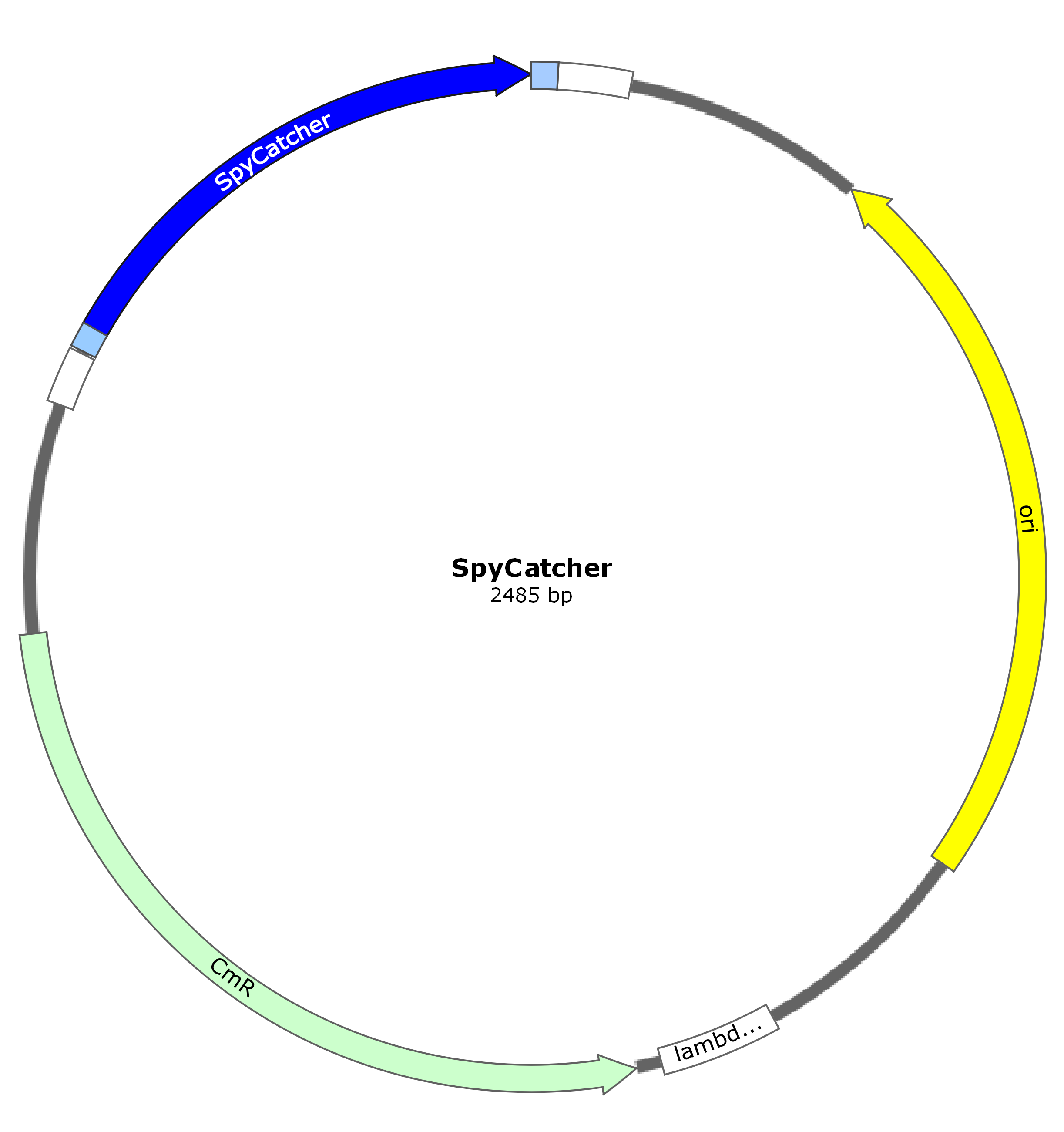
Using BBa_K1321337 (sfGFP in Freiburg format) and BBa_K1650037 (SpyCatcher), we make the fusion protein sfGFP-SpyCatcher to be expressed on the gas vesicle surface after fusion to GvpC.
Choice of BioBricks
BBa_K1321337 (sfGFP in Freiburg format) was chosen because superfolder GFP exhibits intense bright green fluorescence (making it easy to assay) and folds easily into an extremely stable structure — its half-life for unfolding at room temperature is estimated at 28 years! BBa_K1650037 (SpyCatcher) was chosen as it possesses a 6xHis tag near the N-terminus, which allows the fusion protein to be easily purified using a Ni-NTA column.


Our Modification — BBa_K2319002 (GGSGSGSS linker)
Between the sfGFP and SpyCatcher protein domains, we have inserted a short (8 aa), flexible, hydrophilic linker comprising Gly and Ser residues that allows sfGFP and SpyCatcher to fold independently of each other. Serine, like threonine, has a hydroxyl group that contributes to stability by interacting with the aqueous cellular environment. Glycine, again due to its small size, maintains flexibility of the linker.
This linker region also contains a BamHI restriction site (G\GATCC) whose in-frame translation is Gly-Ser. This BamHI site is used to link these two protein domains during assembly of the fusion construct.
Designing a fusion protein: mCherry-SpyTag


