Jol-Fengzi (Talk | contribs) |
|||
| (23 intermediate revisions by 4 users not shown) | |||
| Line 3: | Line 3: | ||
{{:Team:SUSTech_Shenzhen/themeCss}}<!--------整体布局,勿动-------------------------------> | {{:Team:SUSTech_Shenzhen/themeCss}}<!--------整体布局,勿动-------------------------------> | ||
| − | {{:Team: | + | {{:Team:SUSTech_Shenzhen/templates/page-header-part|title=Characterization|subtitle=Parts}} |
| − | + | ||
{{:Team:SUSTech_Shenzhen/main-content-begin}}<!---------内容的样式----------------> | {{:Team:SUSTech_Shenzhen/main-content-begin}}<!---------内容的样式----------------> | ||
| − | |||
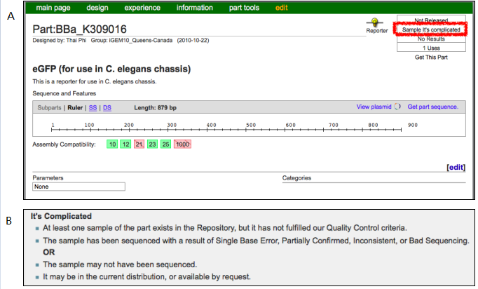
| − | + | We tried to utilize GFP from biobricks distribution to genetically label neuron AWB in C. elegans’ (Caenorhabditis elegans) head. The biobricks part that specifically marked for C. elegans is the <html><a href="http://parts.igem.org/Part:BBa_K309016">BBa_K309016</a></html> submitted by iGEM10_Queens-Canada . Unfortunately, the sample status shows that ”it’s complicated”, which means the sample may not have been sequenced or there is some problems on it (Figure 1). | |
| − | {{SUSTech_Image_Center_fill-width | filename=T--SUSTech_Shenzhen-- | + | {{SUSTech_Image_Center_fill-width | filename=T--SUSTech_Shenzhen--Parts-contribution.png|width=1000px|caption=<B>Figure 1. Parts information on BBa_K309016. A)</B> The part registry page for BBA_K309016; <B>B)</B>:Description of ‘It’s Complicated’ in Registry.}} |
| + | To use GFP for <i>C.elegans</i>, we needed to modify and confirm this GFP. After getting sequence of <html><a href="http://parts.igem.org/Part:BBa_K309016">BBa_K309016</a></html> sequence, we found that the part doesn’t start with start codon “ATG". We optimized it by deleting 9bp in front of the start codon. Then the coding sequence starts with “ATG”(Figure 2). | ||
| + | {{SUSTech_Image_Center_8| filename=T--SUSTech_Shenzhen--Charicterization2.png|width=1000px|caption=<B>Figure 2. Alignment between <html><a href="http://parts.igem.org/Part:BBa_K309016">BBa_K309016</a></html> and <html><a href="http://parts.igem.org/Part:BBa_K2492000">BBa_K2492000</a></html> (optimized GFP) at start codon.</B>}} | ||
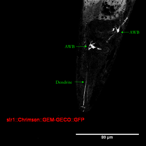
| − | + | We successfully improved characterization of GFP for <i>C.elegans</i>. We submit this optimized GFP as our part <html><a href="http://parts.igem.org/Part:BBa_K2492000">BBa_K2492000</a></html>. We constructed a Str-1::Chrimson::GEM-GECO::GFP fusion gene and injected it into <i>C.elegans</i>. Promoter Str-1 drives the expression of GFP in neuron AWB, which indicate the AWB location. | |
| − | We | + | We capture the image of GFP specifically expressed in AWB with confocal microscope and clearly observe AWB with its dendrite(Figure.3). |
| − | + | {{SUSTech_Image_Center_8 | filename=T--SUSTech_Shenzhen--Parts-Charcterization2.png|width=1000px|caption=<B>Figure.3 Head of <i>C.elegans</i></B>}} | |
| − | + | ||
| − | + | ||
| − | + | ||
| − | + | ||
| − | {{SUSTech_Image_Center_8 | filename=T--SUSTech_Shenzhen--Parts-Charcterization2.png|width=1000px|caption=<B>Head of <i>C.elegans</i></B>}} | + | |
Latest revision as of 02:04, 15 December 2017
Characterization
Parts
We tried to utilize GFP from biobricks distribution to genetically label neuron AWB in C. elegans’ (Caenorhabditis elegans) head. The biobricks part that specifically marked for C. elegans is the BBa_K309016 submitted by iGEM10_Queens-Canada . Unfortunately, the sample status shows that ”it’s complicated”, which means the sample may not have been sequenced or there is some problems on it (Figure 1).
To use GFP for C.elegans, we needed to modify and confirm this GFP. After getting sequence of BBa_K309016 sequence, we found that the part doesn’t start with start codon “ATG". We optimized it by deleting 9bp in front of the start codon. Then the coding sequence starts with “ATG”(Figure 2).

We successfully improved characterization of GFP for C.elegans. We submit this optimized GFP as our part BBa_K2492000. We constructed a Str-1::Chrimson::GEM-GECO::GFP fusion gene and injected it into C.elegans. Promoter Str-1 drives the expression of GFP in neuron AWB, which indicate the AWB location.
We capture the image of GFP specifically expressed in AWB with confocal microscope and clearly observe AWB with its dendrite(Figure.3).


