| Line 24: | Line 24: | ||
#myScrollspy ul li ul li a { | #myScrollspy ul li ul li a { | ||
| − | + | margin-left:60px; | |
} | } | ||
</style> | </style> | ||
Revision as of 16:21, 1 November 2017
Notebook
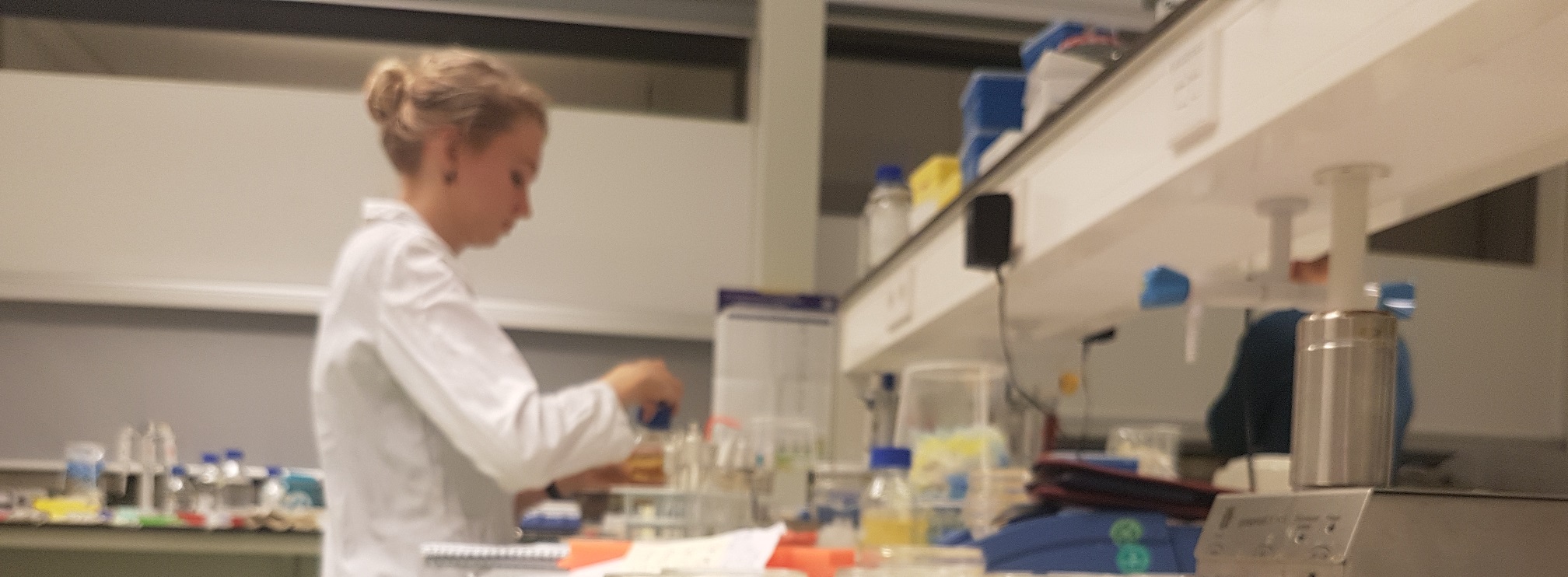
Here you will find our complete labjournal. We documented our daily lab work per week.
Week 23
Although we obtained the keys of our lab on the first of June we have not yet performed any real experiments. The entire week was spent on setting up the lab, e.g. getting all materials and machines needed. Also we made some media and standard buffers and we have produced our first batch of competent E. coli DH5α at the end of this week.
Week 24
General
This week we could finally start doing some real experiments in the lab! We started with optimizing the heat-shock time in the transformation protocol. We did this by transforming our competent cells with DNA from the competent cell test kit with various heat shock times (0-60 seconds). We found that the highest efficiency was obtained when using a heat shock of 30 seconds.Interlab
Because we did not have fully finished the design of any of the experiments for our own project yet, we started with the Interlab study. As a result we can proudly say that we were the first team to complete the Interlab project this year!!We have successfully transformed all test devices of the interlab studies. Of each of the transformation plates one colony was picked, which was inoculated and grown overnight. After plasmid isolation of the ON cultures restriction analysis with EcoRI showed that all colonies contained the correct plasmids. In the remainder of the week we have completed the measurements according the plate reader protocol. More information about the interlab studies can be found on our interlab page.
This and all subsequent experiments requiring plate reading/GFP/RFP were performed with Synergy Mx.Week 25
General
We have inoculated one of the red colonies from the heatshock-test and isolated the plasmid (pSB1C3:bba_J04450) from it. This plasmid was used to confirm that the restriction enzymes, which were left over from the previous iGEM team of Groningen, worked properly. Moreover this plasmid was also used for cloning parts into the pSB1C3 backbone.Lactis toolbox
After completing the interlab we have worked on the lactis-toolbox. We started with two different parts of this sub-project. First was the preparation of electrocompetent L. lactis NZ9000 cells and optimization of the transformation protocols for it. The second step was the preparation of a pTet-GFP/RFP and a pLac-GFP/RFP construct to test these promoters in L. lactis. From next week progress for this part of the project will be shown under the header ‘promoter testing’. This week we produced our first batch of competent lactis cells and determined a growth curve for NZ9000 in SGM17+glycine medium (see figure below). For the second part we successfully transformed E. coli with pLac (bba_), pTet (bba_), sfGFP (bba_) and a RBS (bba_). Insert figure lactis growth curveWeek 26
dCas9
We have tried to transform our E. coli with the dCas9-Omega fusion protein several times, however all attempts failed until we had no DNA left over. To solve this problem we determined to use the pJWV-102-PL-dCas9 and make the dCas9 biobrick compatible ourselves.Spacer acquisition
We obtained the gDNA of a clinical isolate of S. pyogenes from J.M. van Dijl. We tried to PCR out the entire CRISPR operon at once using primers (G1-G4) failed. We have tried the PCR several time including with a temperature gradient PCR. But since all attempts failed we think this isolate may have not contained the CRISPR system. The problem was solved when the plasmids of the Heler lab arrived (see next week).Lactis toolbox
We have obtained five L. lactis vectors from our supervisor H. Karsens is not in the supervisors list in different host strains (see Table below). The cultures were grown overnight and the plasmids were isolated successfully. table from driveWeek 27
Spacer acquisition
This week the Heler plasmids arrived so we could start with creating the different DNA fragments for the gibson assembly of the hCas9 operon.We started with PCR amplifying the DNA fragments from the pWJ40 plasmid using the primer pairs G19, G20 and G23, G24. The resulting PCR products were treated with DpnI and cleaned with a PCR clean up kit.Lactis toolbox
The competent lactis cells that were produced in week 25, were tested by transforming them with the isolated pNZ8048 plasmid. The transformation did not work out, probably because the Chloramphenicol concentration in the plates was too high (25ug/ml). Also the pNZ8048 plasmid was linearized and the biobrick prefix and suffix were incorporated. The PCR was performed using Phusion polymerase and primers … (gel?)The sfGFP was cloned into the linearized vector using restriction ligation with EcoRI and PstI. The linearized pNZ8048 was not digested with DpnI, because L. lactis plasmid DNA is not methylated and therefore DpnI cannot digest the template. The ligation product was stored in the fridge.
Promotor testing
For this part we successfully transformed and isolated bba_, which contains the RFP plasmid. Also we completed the first round of 3A assemblies, shown in the table below. table from drive The plates for pLac_GFP and pLac_RFP contained four colonies and the pTet_RBS plate contained only one colonies. All colonies were heavily overgrown since they were left at 37C for the entire weekend.Week 28
General
We finished the first part of the design of the dCas9 and the spacer acquisition projects and we ordered the second batch of primers (G15-G37). New competent E. coli DH5α and lactis cells were produced. Add more infodCas9
From our supervisor C. Huang we received E.coli DH5α containing pJWV-102-PL-dCas9. These cells were plated and one colony was picked and inoculated. The plasmid was isolated from the ON cultures and analysed by restriction analysis with PstI (see gel next week).Promotor testing
The colonies of the 3A assemblies that were heavily overgrown were re-plated. Each plate contained many colonies and those on the pLac-GFP plate were coloured green. Restriction analysis of isolated plasmids from one of each of the colonies showed the correct size for pLac_GFP and pTet_RBS, but not for pLac_RFP. Since we only wanted to use the constructs for the function of pLac in lactis and we only need one marker for this we decided to not retry the pLac_RFP assembly.Week 29
General
This week the second batch of primers arrived, so we could finally continue our work in the dCas9 and the spacer acquisition projects. PCR amplification of gBLOCKS?Spacer acquisition
This week the gBlocks arrived. The I473F/EcorI gblock was amplified using the primer pair G21, G22. The other gBlock contained both the tracer RNA and a part of the hCas9 operon. It was first inserted into pSB1C3 by restriction ligation with EcorI and PstI and transformed into E. coli DH5α. The transformation went well and the plasmids checked by a restriction analysis with EcorI. We planned to seperate the tracer RNA from the hCas9 operon when we linearized the plasmid using PCR. With the primer pair G27, G28 we wanted to create a linearized tracerRNA pSB1C3 vector and with primer pair G17, G18 we wanted to create a linearized hCas9 vector, which would be the backbone for the gibson assembly. PCR 1,2 ? using heller plasmids?dCas9
The isolated pJWV-102-PL-dCas9 was used to PCR out the dCas9 gene using primers G35 and G36. Since it is a large size construct (4,2 kb) we used Phusion polymerase and a Tm of ... . At the moment we thought that we had the correct PCR product, because we did not analyse the gel carefully enough. In the figure below the PCR fragment is shown together with the restricted pJWV-102-PL-dCas9 and of pSB1C3:dCas9 (see next week). From the gel it can be seen that the actual product sizes on the left are larger than they should be. On the right side of the gel the same products are shown, but this time with the correct plasmid (see …). Redo gel: dCas9 old vs new plus uitleg in onderschrift (grootte)Week 30
Spacer acquisition
Trouble shooting PCR for gBlock separation of hCas9-tracer RNA with different annealing temperatures and addition of DMSO to the PCR mix.dCas9
As said in the report of last week the PCR fragment was cloned into [SB1C3:GFP (bba_) via restriction ligation with XbaI and PstI. The transformation resulted in four colonies of which the plasmids were isolated and analyzed by restriction with PstI and EcorI. The results are shown in the figure below on the right. Colonies 2 and 3 seemed to have bands of the right size and those plasmids were used for follow-up experiments. However as explained in the previous report this dCas9 turned out to be wrong, so all other experiments using this part failed. GEL 28-07 colnyPCR+restrictionanalysisGEL (M|four hCas9 colonies|four dCas9 colonies|M)Week 31
General
gBlock dCas9 VRER and hCas9I473V were ligated into linearized pJET1.2 bluntend vector via blunt end ligation. The pJET vector was a gift from our supervisor C. Pohl, who also gave us two primers pJet_Fwd and pJet_Rev which bind on both the pJet backbone and the pSB1C3 backbone. These primers were used for sequencing and to confirm whether a fragment was inserted into pSB1C3.Lactis toolbox
Transformation of lactis with pNZ8048. The transformation product was plated on plates containing different concentrations of chloramphenicol (0-15 ug/ml). 5 ug/ml was found to be the best concentration. Moreover we linearized the pIL252 and pIL 253 backbones using Phusion polymerase with primers G25, G26.Spacer acquisition
To separate the TracrRNA part and the prefix-suffix part from the plasmid containing the hCas9 gblock we wanted to do two PCRs. One to obtain the pSB1C3:pUsp45TracrRNA and one with the pSB1C3 containing the part used for the hCas9-operon. However this week all attempts of performing this PCR failed.Week 32
General
New primers for dCas9 Quickchange and separation of hCas9 combi gblock were ordered.This week we succesfully transformed E. Coli with the pJET vectors containing the gBlocks. The colonies on the plate were analysed by colony PCR with the pJet primers and for both pJET-constructs we isolated the plasmid of the colonies with a positive size of the insert. Gel 9/8?
Lactis toolbox
To optimize the transformation protocol of lactis further we tested different DNA to cell ratio’s. We used either 50 or 100 ul of competent lactis cells and transformed them with 50, 100 or 250 ng of pNZ8048. The volume of DNA that was added was respectively 0.5, 1 or 2,5 ul. The results are shown in the table below. Although we find it strange that using more cells did not result in more colonies the result does show that adding more DNA does not result in more colonies. We think that this is not caused by the amount of DNA that is added, but by the volume of the DNA that is added. Adding a larger volume will dilute the competent cells further, which will lead to a decreased transformation efficiency.Spacer acquisition
After testing even more PCR conditions we finally thought that we succeeded in creating the pSB1C3 containing the tracer RNA part. The PCR which succeeded was a temperature gradient PCR using phusion polymerase, primers G27, G28 and a annealing temperature ranging from 55-65 C. After the PCR the product was THIJS work up for pUsp45 TracrRNA. table from driveWeek 33
General
Since the PCR’s for the quickchange PCR of dCas9 and also the PCR of the gblock of hCas9 kept failing we designed and ordered new primers for them. (primers … - … were ordered). Also we send the isolated pJET-gblock plasmids and pSB1C3 containing the hCas9 combigblock. RESULT?dCas9
Besides ordering the new quickchange primers we also rechecked our template DNA together with the PCR fragment and the original dCas9 vector. At this point we found out that the dCas9 PCR might be too big. MART15-08 sequencing results of three samples (other samples did not work out)Week 34
Spacer acquisition
The pUSP45 tracer RNA(see week August 07-13) was put into an iGEM backbone and transformed into E. coli DH5α according to protocol. There were colonies visible on the plates and these were used for ON cultures. The plasmids were isolated and send for sequencing.dCas9
Since the PCR product of dCas9 seemed larger than the expected size we redid the PCR to isolate dCas9 from the pJWV-102-PL-dcas9 plasmid. However the product was the same size as the one in the previous experiment.Week 35
Week 36
Week 37
Week 38
Week 39
Week 40
Week 41
Week 42
Week 43
Week 44
Lactis toolbox
After constructing the promoter biobricks and expression vectors, the pNisA promoter was validated in a similar fashion as the cells of the InterLab study were examined. After growing an overnight culture at 30°C, the cells were diluted to an optical denisty of 0.1 in fresh GM17 medium. When the optical density reached a value between 0.5 and 0.6, the culture was split into 5 portions and induced with various amounts of nisin. The used concentrations were 1 ng/ul, 10 ng/ul, 45 ng/ul and 80 ng/ul and were administered from at stock dissolved in 10% glucose. The cells were grown for 6 hours at 30°C and a sample was taken every 45 minutes. The samples were stored in the dark and on ice until further use. Once all samples were collected, the cells were havested by centirfugation and resuspended in chemically defined medium. The cell suspensions were analysed by two measurements. The first was an absorbance measurement at 600 nm to determine the optical density and therfore the amount of cells present. The second measurement determined the amount of expressed GFP by exciting at 489 nm and measuring emission at 512 nm.What should this page have?
- Chronological notes of what your team is doing.
- Brief descriptions of daily important events.
- Pictures of your progress.
- Mention who participated in what task.
Inspiration
You can see what others teams have done to organize their notes:


